Book on Whatsapp
9892101616

Embolization Offers Minimally Invasive Approach to Enlarged Prostate Management
Radiology
Sat Jun 08 2024
Researchers have identified a promising minimally invasive procedure for treating benign prostatic hyperplasia (BPH). It is commonly known as enlarged prostate.
BPH is a widespread condition affecting older men. The condition leads to bothersome urinary symptoms like:
- Weak stream
- Dribbling at end of urination
- Increased frequency and of urgency of urination
- Frequent urination at night.
Some men experience mild symptoms. Others face a significant decline in quality of life. They struggle to sleep through the night and limit their daily activities due to constant bathroom urgency.
Traditional BPH treatments may have limitations. Surgical procedures are effective. But they carry a risk of complications. Side effects may include incontinence and erectile dysfunction. Medications may not be effective for all patients. Some men may experience unwanted side effects that further impact their quality of life.
Prostate artery embolization (PAE) emerges as a possible alternative. This minimally invasive procedure is performed by specially trained interventional radiologists. It involves inserting a thin catheter through the wrist and guiding it using real-time X-ray imaging to the arteries supplying blood to the prostate. Tiny microscopic beads are then deployed through the catheter to block blood flow. It causes the prostate to shrink over time.
PAE- Is It Safe?
Initial studies suggest PAE is both safe and effective. Men who have undergone PAE report substantial improvement in urinary symptoms. Early data shows significant reductions in nighttime urination and a stronger urinary stream. It allows men to regain control of their bladder function.
PAE offers several advantages over traditional BPH treatments:
- Minimally invasive approach: Unlike surgery, PAE requires only a small incision in the wrist, minimizing recovery time and discomfort.
- Low complication risk: PAE is associated with a lower risk of complications compared to surgery.
- Negligible risk of incontinence or erectile dysfunction: Unlike some BPH treatments, PAE is designed to spare nerves and blood vessels responsible for sexual function and urinary control.
- Expedited recovery time: Following PAE, most men recover quickly and can resume normal activities within a day or two.
While PAE is a relatively new procedure, long-term data is still being collected. However, for men with bothersome BPH who are not suitable candidates for surgery or medication, PAE presents a potentially life-altering option.
Men experiencing urinary symptoms consistent with BPH should consult their physicians. A simple urine flow test can be used to assess urinary flow rate and identify potential BPH. Early diagnosis and treatment can help prevent complications and improve quality of life.
The availability of diverse treatment options empowers men with enlarged prostate to take an active role in their healthcare decisions. By discussing all available options with their physicians, men with BPH can choose the treatment approach that best suits their individual needs and preferences, allowing them to regain control of their urinary symptoms and improve their overall well-being.
Related Tests
Related Packages

Food Allergy Profile
Food Allergy Profile Test for Understanding Food Sensitivities
The food allergy profile test is a comprehensive diagnostic tool designed to identify specific food allergens that may be triggering allergic reactions in your body. Common symptoms of food allergies include rashes, digestive issues, difficulty breathing, and swelling. Identifying the cause of these reactions is essential for effective management and prevention of further complications.
At Manipal TRUtest, the food allergy panel test price is affordable, ensuring everyone has access to accurate allergy testing. This test evaluates your body's immune response to various foods by detecting specific Immunoglobulin E (IgE) antibodies. It covers a wide range of allergens, including dairy, nuts, seafood, grains, and fruits, making it an essential tool for individuals experiencing unexplained allergic reactions.
Booking your food allergy profile test with Manipal TRUtest is simple and hassle-free. We also offer convenient at-home testing services, where a skilled phlebotomist collects your blood sample at your residence. Results are delivered quickly, allowing you to take timely action.
Understanding your food allergies can significantly improve your quality of life. Take charge of your health by scheduling the food allergy panel test today at Manipal TRUtest and enjoy transparent pricing and precise results tailored to your needs.
₹ 11200

TRU Fit Package
₹ 4499
Related Blogs

Radiology
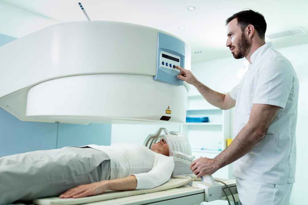
Everything You Should Know About Head MRI and Cost
Magnetic Resonance Imaging (MRI) is a powerful diagnostic tool that helps medical professionals visualize the internal structures of the body in detail. A Head MRI is a non-invasive imaging procedure that creates detailed images of the brain, skull, blood vessels, and surrounding tissues. It is widely used to detect and evaluate various neurological conditions, injuries, and abnormalities.
What is a Head MRI?
A Head MRI uses strong magnetic fields and radio waves to generate images of the brain and other structures inside the head. Unlike X-rays or CT scans, it does not involve ionizing radiation, making it safer, especially for repeated use. The procedure usually takes around 30 to 60 minutes and may involve the use of contrast dye (gadolinium) to enhance the visibility of certain areas.
Why is a Head MRI Recommended?
Doctors recommend a head MRI for various reasons, including:
- Persistent Headaches or Migraines: To rule out underlying neurological causes.
- Seizures: To identify any abnormalities in the brain tissue.
- Stroke Symptoms: To detect signs of stroke or mini-strokes (TIAs).
- Traumatic Brain Injury: To assess the extent of damage following an accident or injury.
- Brain Tumors: To locate and evaluate tumors or cysts.
- Multiple Sclerosis (MS): To detect and monitor lesions or inflammation in the brain.
- Infections: Such as encephalitis or meningitis.
- Dizziness or Vision Problems: When symptoms point to a neurological cause.
How is a Head MRI Performed?
The patient lies on a movable table that slides into the MRI scanner a large, cylindrical machine. During the scan, it is crucial to stay still to obtain clear images. The procedure is painless, although the machine may make loud thumping or tapping sounds. Patients with claustrophobia may be given a mild sedative. If contrast is required, it is injected through a vein in the arm before or during the scan.
Safety Considerations
While MRIs are generally safe, they are not suitable for everyone. Individuals with pacemakers, metal implants, or certain other medical devices may not be eligible for an MRI. It’s essential to inform the radiologist about any medical conditions, allergies, or pregnancy before the procedure.
Cost of a Head MRI in India
The cost of a head MRI can vary significantly based on location, the type of facility (private vs. public), and whether contrast dye is used. Some factors influencing the cost include:
- City or Region: Tier 1 cities usually have higher costs.
- Hospital or Diagnostic Center: Premium centers may charge more.
- Type of MRI Machine: High-resolution 3 Tesla MRIs may cost more than standard 1.5 Tesla machines.
- Insurance Coverage: Some health insurance policies cover the cost of diagnostic tests, including MRIs.
A Head MRI is an invaluable tool in modern medicine, offering detailed insights into the brain and related structures. While the procedure is safe and non-invasive, it should only be performed upon a doctor’s recommendation. Knowing the approximate cost and process helps patients make informed decisions about their health and diagnostics.
If you are experiencing symptoms such as persistent headaches, memory loss, or neurological changes, consult your healthcare provider. Timely imaging can lead to early diagnosis and better treatment outcomes.
For accurate and affordable MRI scans , visit the nearest Manipal TRUtest center
Radiology
The Importance of MRI Scans in Detecting Health Issues
Medical imaging has transformed healthcare, and MRI (Magnetic Resonance Imaging) stands out as one of the most important tools for early and accurate diagnosis. An MRI scan uses powerful magnets and radio waves to create detailed images of the inside of the body, without the use of radiation. This helps doctors identify health problems quickly and plan the best treatment for their patients.
What Makes MRI Scans So Valuable?
Unlike X-rays or CT scans, MRI provides highly detailed pictures of soft tissues, organs, and even the nervous system. It is especially useful in detecting issues in the brain, spine, joints, heart, and internal organs. When symptoms are unclear, an MRI can uncover hidden problems that other scans might miss. This makes it a trusted choice for doctors who want a clearer view of what’s happening inside the body.
At Manipal TRUtest, advanced MRI services ensure that patients get accurate and reliable results quickly, helping them start treatment without unnecessary delays.
Common Health Issues Detected by MRI
MRI scans are used to diagnose a wide range of conditions, such as:
- Brain and spinal cord issues: MRI is excellent at detecting brain tumors, strokes, multiple sclerosis, and spinal cord injuries.
- Joint problems: If you have unexplained joint pain, an MRI can show damage to ligaments, cartilage, and muscles.
- Heart conditions: Doctors use MRI to assess the structure and function of the heart, helping to diagnose heart diseases early.
- Organ health: MRI scans can spot liver diseases, kidney problems, and abnormalities in other internal organs.
By offering clear images, MRI helps in creating the right treatment plan, making recovery faster and smoother.
When Should You Consider an MRI?
Doctors usually recommend an MRI when they need a deeper understanding of a health issue. You may need an MRI if:
- There is a need to monitor known conditions like tumors or injuries.
- You experience unexplained symptoms that cannot be diagnosed with other imaging tests.
- You have persistent headaches, seizures, or dizziness.
- You are suffering from chronic joint or back pain.
Choosing a trusted center like Manipal TRUtest for your MRI scan ensures that you are in safe hands, receiving quality care at every step.
How to Prepare for an MRI Scan
Preparing for an MRI scan is usually rather straightforward. You may be asked to remove metal items such as jewellery before the scan. If you have any metal implants or pacemakers, let the technician know beforehand. You may receive a contrast dye to enhance the imaging during the scan. The MRI scan is painless, and you will usually be able to go home shortly thereafter.
At Manipal TRUtest , the medical team guides you through every step, making sure you are comfortable and well-informed before and after the procedure.
The role of MRI scans in modern medical diagnosis is substantial. An MRI scan has the potential to find serious health issues quickly and even save lives. If it is a brain condition, a heart issue, or pain with an unknown origin, an MRI provides the clarity needed to move forward with confidence. Why not trust a reliable service to perform the MRI? Take the time to be proactive with your health, and go to Manipal TRUtest for accurate MRI imaging.
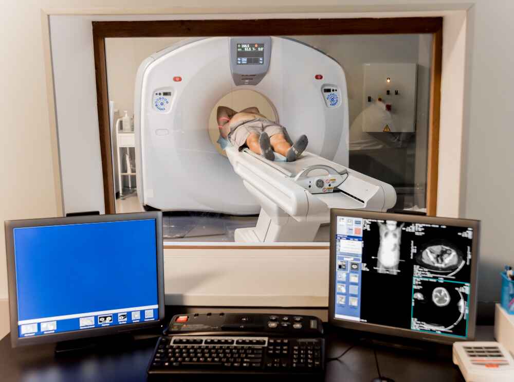
Radiology
When and Why You Might Need an Abdominal Scan
The abdomen is where important organs such as the liver, kidneys, pancreas, gallbladder, and intestines all reside. When something doesn't feel right in this area, doctors often recommend an abdominal scan to figure out what is going on inside the abdomen. These scans are safe, quick, and can help provide important information for the next step in your path to better health.
What is an Abdominal Scan?
An abdominal scan is an imaging test that allows doctors to view organs and tissues inside the belly. It can be done by different procedures such as ultrasound, CT scan and MRI. Each technique has its own benefits, depending on what the doctor is looking for. The scans are quick and painless, and can sometimes identify problems early on before they have developed into symptoms.
When Might You Need an Abdominal Scan?
There are many reasons a doctor might recommend an abdominal scan. Some of the most common situations include:
1. Unexplained Pain
If you are experiencing pain in your abdomen that does not go away, a scan can help identify the cause. It could be due to gallstones, liver issues, infections or kidney stones,
2. Digestive Problems
Persistent issues like nausea, vomiting , and changes in bowel habits may require a closer look. An abdominal scan can reveal if there are any blockages, swelling, orabnormalities in the digestive organs.
3. Injury or Trauma
After an accident or a fall, an abdominal scan is often done to check for internal bleeding and organ damage. Even if there are no external injuries, it is important to ensure that the internal organs are safe.
4. Suspected Infections
Infections in the abdomen, such as a reddened appendix or an abscess, which can be dangerous if not treated quickly. A scan helps detect these circumstances so that the right treatment can begin without delay.
5. Monitoring Existing Conditions
For people already diagnosed with conditions like liver disease, kidney problems, and cancer, regular abdominal scans are important. They help monitor the disease’s progress and assess how well the treatment is working.
6. Checking for Tumors or Growths
An abdominal scan can spot abnormal growths like tumors, lumps, and cysts. Early detection makes it easier to manage these conditions effectively.
Different Types of Abdominal Scans
Depending on the situation, your doctor may choose one of the following scans:
- Ultrasound: Common for checking gallbladder, liver, and kidneys. It’s quick and uses sound waves instead of radiation.
- CT Scan: Provides more detailed images and is often used to spot injuries, infections, or tumors.
- MRI Scan: Gives very detailed pictures, especially useful for soft tissues and blood vessels.
Each scan has its own benefits, and your healthcare provider will decide which one suits your needs best.
How to Prepare for an Abdominal Scan
Preparation depends on the type of scan. For ultrasounds, you might be asked to fast for a few hours beforehand. For CT or MRI scans, you may need to drink a contrast liquid or receive an injection to make certain areas clearer. Always follow the instructions given by the healthcare team to ensure accurate results.
An abdominal scan is a powerful tool that helps doctors see inside your body without surgery. Whether it’s unexplained pain, digestive troubles, or simply monitoring an ongoing condition, these scans play a key role in keeping you healthy. If your doctor recommends an abdominal scan, it is a step towards understanding your health better and taking timely action when needed.

