Book on Whatsapp
9892101616

How To Test Yourself For COPD
Lungs
Fri Nov 17 2023
If you suspect you may have chronic obstructive pulmonary disease (COPD), it is essential to consult a healthcare professional for an accurate diagnosis and proper evaluation. While there are no definitive at-home tests for COPD, you can perform some preliminary assessments to determine if you have symptoms consistent with the condition. Here are a few steps you can take:
1. Self-Assessment: Evaluate your symptoms. Common indications of COPD include:
- Chronic coughing, often with excessive mucus production
- Shortness of breath, especially during physical activity
- Wheezing or a whistling sound when you breathe
- Chest tightness or discomfort
2. Medical History: Review your medical history to identify potential risk factors associated with COPD. These include:
- Smoking history: COPD is primarily linked to smoking, both active and passive.
- Occupational exposure: Certain workplace environments can increase the risk, such as exposure to chemicals, dust, or fumes. Read More : - Unveiling the Detrimental Effects: The Hazardous Impact of Smoking on Human Health
- Family history: COPD may have a genetic component, so a family history of the condition could increase your risk.
3. Spirometry Test: Spirometry is a lung function test commonly used to diagnose and assess COPD. It measures how much air you can exhale forcibly and how quickly you can do so. This test is performed at a medical facility and requires specialized equipment.
4. Consult a Healthcare Professional: If you suspect you have COPD based on your symptoms and risk factors, make an appointment with your healthcare provider. They will be able to evaluate your condition more accurately through a physical examination, review of your medical history, and potentially order further diagnostic tests, including spirometry.
5. Physical Examination: During a physical examination, your healthcare provider may listen to your lungs with a stethoscope to check for abnormal breath sounds, such as wheezing. They might also examine your chest and ask about your symptoms in detail.
6. Chest X-Ray: A Chest X-ray can help rule out other conditions that may have similar symptoms to COPD, such as lung infections or heart problems. It can also identify any structural abnormalities in your lungs.
7. Blood Tests: While there are no specific Blood Tests to diagnose COPD, blood tests can help evaluate your overall health and identify any underlying conditions that might be contributing to your symptoms. For example, blood tests can assess oxygen levels, check for signs of infection, or measure inflammatory markers.
8. Pulmonary Function Tests (PFTs): Pulmonary function tests evaluate how well your lungs are functioning and are often used to diagnose and monitor COPD. These tests measure lung volume, airflow, and gas exchange. The most common test is spirometry, which assesses how much air you can forcefully exhale after taking a deep breath.
9. Arterial Blood Gas (ABG) Test: An ABG test involves drawing blood from an artery, typically from the wrist, to measure the oxygen and carbon dioxide levels in your blood. This test determines how well your lungs are oxygenating your blood and if you have any abnormalities in gas exchange.
It's important to note that only healthcare professionals can diagnose COPD definitively. Self-assessment and preliminary evaluations can give you an idea of your symptoms and risk factors, but consulting with a healthcare provider is necessary for an accurate diagnosis and appropriate management plan.
Remember, self-assessment and preliminary evaluations are not a substitute for professional medical advice. Seeking guidance from a healthcare professional is crucial for an accurate diagnosis and appropriate management of COPD or any other health condition.
Manipal TRUtest offers a wide range of diagnostic services with assured quality, accuracy, and trust backed by 70 years of Manipal legacy. You can easily schedule a blood test through our WhatsApp Chatbot, Mobile App, or Website. We also provide the convenience of a Home Sample Collection, where a highly qualified phlebotomist will visit you in the comfort of your own home.
OUR PRESENCE
Blood Test Center in Hyderabad / Blood Test Center in Kolkata / Blood Test Center in Vizag / Blood Test Center in Mumbai / Blood Test Center in Ghatkopar / Blood Test Center in Kolhapur / Blood Test Center in Pune / Blood Test Center in Solapur / Blood Test Center in Rohtak / Blood Test Center in Indore / Blood Test Center in Gurugram / Blood Test Center in Ghaziabad/ Blood Test Center in Bangalore / Blood Test Center in Nashik / Blood Test Center in Nagpur
Related Blogs

Lungs
Lung Cancer Symptoms Causes and Prevention
Lung cancer is one of the most prevalent and life-threatening malignancies worldwide, claiming millions of lives each year. It develops when abnormal cells in the lung tissue grow uncontrollably, forming tumors that impact normal respiratory function. Detecting it early can significantly improve treatment outcomes and survival rates.
Common Symptoms of Lung Cancer
In many cases, the disease progresses silently, with symptoms emerging only in the later stages, making early detection a challenge. Key warning signs may include –
- Cough that worsens over time,
- Coughing up blood or rust-tinged sputum
- Unexplained shortness of breath during routine activities,
- Chest pain that intensifies with deep breathing or coughing, hoarseness, significant
- Unexplained weight loss,
- Chronic fatigue, and recurrent respiratory infections such as bronchitis or pneumonia.
While these symptoms can also indicate other conditions, their persistence warrants prompt medical evaluation.
Causes and Risk Factors
The leading cause of lung cancer is tobacco smoking, accounting for approximately 85–90% of all cases. Cigarette smoke contains thousands of harmful chemicals, many of which are known carcinogens that damage lung tissue at the cellular level. However, non-smokers are not immune to the disease. Long-term exposure to second-hand smoke, inhalation of occupational carcinogens such as asbestos, radon gas, and diesel exhaust, prolonged exposure to polluted air, a family history of lung cancer, and pre-existing lung conditions like chronic obstructive pulmonary disease (COPD) or pulmonary fibrosis can all significantly elevate the risk. These factors cause genetic mutations within lung cells, triggering abnormal growth and tumor formation.
Prevention Tips
Although not all cases can be prevented, adopting healthier lifestyle choices can greatly reduce the likelihood of developing lung cancer. Quitting smoking remains the single most effective preventive measure, with benefits beginning almost immediately after cessation. Avoiding environments with second-hand smoke, testing homes for radon gas, using protective equipment when working around hazardous substances, and improving indoor air quality are also essential. A nutrient-rich diet abundant in fruits, vegetables, and antioxidants, coupled with regular physical activity, can strengthen the immune system and support overall lung health. For individuals at high risk, particularly long-term smokers, annual low-dose CT scans can facilitate early detection, improving treatment outcomes.
In essence, lung cancer is a formidable global health concern, but increased awareness, timely medical attention, and proactive preventive strategies can save countless lives.
By recognizing symptoms early, understanding the underlying causes, and embracing healthy habits, individuals can take meaningful steps toward safeguarding their respiratory health and enhancing their quality of life.
If you experience persistent respiratory symptoms, don’t ignore them. Book your screening with Manipal TRUtest and take proactive steps toward safeguarding your lung health.

Lungs
The Benefits of Routine X-Rays for Bone and Lung Health
Modern medicine relies heavily on X-rays as a diagnostic tool since they provide important information about the state of the lungs and bones. When utilised properly, routine X-rays can help identify possible health problems early on, allowing for prompt treatment and improved results. The advantages of routine X-rays for preserving lung and bone health are examined in this article.
1. Early Detection of Bone Conditions
Early detection of bone abnormalities is made possible by routine X-rays. Without imaging, conditions like osteoporosis, fractures, infections, and bone tumours can frequently go undetected. Doctors can diagnose issues before they get worse by using X-rays to see changes in bone density, shape, and alignment.
Routine X-rays can help monitor bone density and identify indications of fractures or bone thinning in people with osteoporosis, a disorder that weakens bones. When osteoporosis is detected early by X-rays, preventive treatments like medication or lifestyle modifications may help slow down bone loss and lower the chance of fracture.
2. Diagnosing Lung Diseases
X-rays can identify several respiratory disorders, making them equally vital for lung health. One of the best methods for identifying conditions like lung cancer, pneumonia, TB, and chronic obstructive pulmonary disease (COPD) is a chest X-ray. Routine chest X-rays can detect early indications of lung issues, even before symptoms manifest, in people with a history of smoking or lung illness.
Abnormalities including fluid accumulation, tumours, or infection symptoms can be shown on chest X-rays. For example, an X-ray can reveal inflammatory lung regions in pneumonia cases, enabling timely treatment. X-rays can identify worrisome growths in the case of lung cancer that may need more testing, like a biopsy or CT scan.
3. Monitoring Chronic Conditions
Additionally, routine X-rays are crucial for tracking long-term illnesses. Regular X-rays may be necessary for patients with chronic conditions like COPD, TB, or asthma in order to monitor changes in lung function and spot any problems. Doctors can avoid additional deterioration and modify treatment methods by identifying changes over time.
People who have had joint replacement surgery or who have persistent arthritis may require routine X-rays to check the condition of their bones and joints. These imaging findings can be used to assess if the bones are worn down or damaged, allowing for prompt treatment to prevent more serious issues.
4. Assessing Injury Recovery
Routine X-rays are frequently performed to evaluate healing following a bone injury. For instance, follow-up X-rays could be necessary for a fractured bone to make sure it is healing properly and to look for any complications like infection or misalignment. Using X-rays to track recovery aids physicians in determining whether more care or modifications are required.
Similarly, X-rays can monitor recovery and make sure the lungs are healing properly in situations when lung function has been reduced by an injury or surgery.
5. Preventive Care and Peace of Mind
Apart from their diagnostic utility, routine X-rays can also offer comfort. Regular X-rays taken during checkups can reassure patients and medical professionals that there are no serious lung or bone problems. Frequent screenings help guarantee that any issues are identified early, improving health outcomes, especially for people who are more susceptible to lung or bone disorders.
Conclusion
The early detection, tracking, and treatment of lung and bone health depend heavily on routine X-rays. These imaging techniques help medical professionals identify problems early, stop more complications, and direct successful therapies by giving information on the state of the lungs and bones. Frequent X-rays are an essential component of preventive healthcare, providing physicians and patients with a useful tool for preserving long-term health.
Lungs
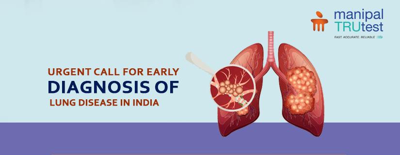
Urgent Call for Early Diagnosis of Lung Disease in India
Health experts are raising alarms about the increasing rates of chronic obstructive pulmonary disease (COPD) in India and the crucial need for early diagnosis and ongoing treatment.
Recent findings have highlighted the urgency of addressing COPD, a serious lung condition that affects a significant portion of the Indian population. The disease is a major health concern, ranking as one of the leading causes of death globally. In India, millions of people are living with COPD, with a substantial percentage suffering from this debilitating condition. Alarmingly, a large number of these individuals are not receiving the necessary treatment.
What is Chronic Obstructive Pulmonary Disease?
COPD is a progressive disease that impairs lung function, making it difficult for affected individuals to breathe. It is commonly associated with smoking, including the use of traditional smoking devices such as bidis and hookahs. The increase in smoking practices among various age groups has contributed to the rise in COPD cases.
The impact of COPD on daily life is profound. The disease not only affects physical health but also diminishes overall quality of life. Despite its seriousness, efforts to identify and treat COPD early are often lacking. Medical professionals emphasize the importance of early diagnosis and treatment to manage the disease effectively and prevent long-term health complications. Without timely medical intervention, COPD can lead to significant health burdens later in life.
The Overview of COPD in India
Experts stress that a significant percentage of COPD cases go undiagnosed in India, highlighting the need for increased awareness and better diagnostic practices. People need regular health checkups for quick diagnosis. Early detection allows for the use of effective medications and lifestyle modifications that can greatly improve patients' quality of life. Simple changes and appropriate treatments can make a significant difference in managing the disease.
COPD generally affects adults over the age of 40. It is primarily caused by smoking, but poor air quality significantly contributes to its prevalence. Both indoor and outdoor air pollution, including particulate matter and gases, exacerbate COPD symptoms and increase hospitalizations. Improving air quality standards is essential for protecting vulnerable populations. An Air Quality Index of over 80, is generally considered to trigger COPD.
One of the key indicators of COPD is an increase in shortness of breath, which signifies changes in lung function. Prompt diagnosis and treatment are crucial to managing the condition and maintaining respiratory health.
How To Detect COPD?
To diagnose Chronic Obstructive Pulmonary Disease (COPD), healthcare providers commonly utilize tests such as spirometry, which measures lung function, and sputum microscopy, which analyzes mucus for signs of infection or inflammation. These tests are essential for confirming the presence and severity of the disease
Conclusion
There is a pressing need for greater awareness and proactive measures to diagnose and treat COPD early in India. By improving public knowledge and encouraging timely medical intervention, it is possible to enhance the quality of life for those affected by this debilitating disease and reduce the overall health impact of COPD.

